ΑΓΓΕΙΟΧΕΙΡΟΥΡΓΟΣ
Επιλεγμένες Επιστημονικές Δημοσιεύσεις
Endovascular Repair of an Inflammatory Abdominal Aortic Aneurysm Combined with a Congenital Pelvic Kidney: Case Report and Literature Review.
Papadoulas S, et al. Aorta (Stamford). 2022 Jun;10(3):135-140. doi: 10.1055/s-0042-1748961. Epub 2022 Nov 1. PMID: 36318935; PMCID: PMC9626033. Department of Vascular Surgery, University of Patras Medical School, Patras, Greece
The coexistence of an abdominal aortic aneurysm and a congenital pelvic kidney is extremely rare. We present a 66-year-old male with an inflammatory aneurysm and an aberrant origin of the superior mesenteric artery. The inflammatory infrarenal abdominal aortic aneurysm with a congenital left pelvic kidney was successfully treated with endovascular repair. Coverage of one out of the two renal ectopic arteries was performed, without clinical evidence of renal function impairment.

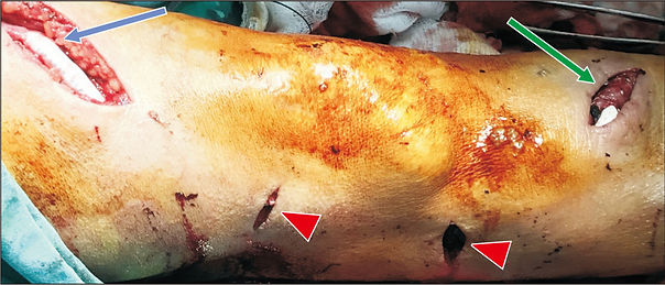
Superficial temporal artery pseudoaneurysm after blunt trauma: A case series
Christos Pitros, Spyros I. Papadoulas et al. Hellenic Journal of Vascular and Endovascular Surgery, Volume 4, Issue 3, 2022. Department of Vascular Surgery, University of Patras Medical School, Patras, Greece
Purpose: To describe our experience with the presentation and management of traumatic superficial temporal artery pseudoaneurysms in our institution. Small case series description: Reviewing all patient’s records during the last 20 years we identified 3 cases with a superficial temporal artery pseudoaneurysm. Two patients had suffered a motorcycle accident and presented with blunt and/or penetrating skin injuries on the head remote of the site of the aneurysm. No helmet was used. The remaining patient presented with skin necrosis on the aneurysm indicating a direct blunt arterial injury after a minor fall. No other severe injuries were apparent. After a diagnostic colour duplex they underwent aneurysm excision and ligation of the superficial temporal artery under local anesthesia. Conclusion: In our patients’ temporal artery pseudoaneurysms were developed after direct arterial transection or traction due to a nearby head injury. Diagnosis was based on clinical examination and confirmed by ultrasound. Treatment consisted of aneurysm excision.

Patient Transfer with Kocher Forceps on the Axillary Artery: A Rare Case of Ongoing Iatrogenic Vascular Injury.
Seretis C, Spyros Papadoulas et al. Vasc Specialist Int. 2022 Mar 31;38:10. doi: 10.5758/vsi.220010. Department of Vascular Surgery, University of Patras Medical School, Patras, Greece
Iatrogenic trauma of the axillary artery by non-vascular surgeons can occur during various general surgical procedures such as resection of soft tissue tumors or axillary lymph node clearance. Prompt recognition, appropriate initial management, and rapid transfer to a tertiary vascular surgery service, if needed, are key steps to ensuring patient safety. Here we present a case of iatrogenic axillary artery injury during the resection of a recurrent soft tissue tumor in a local hospital. The desperate application of a Kocher clamp on the bleeding axillary artery by the operating general surgeons controlled the bleeding but led to further arterial damage. The patient was transferred to our tertiary hospital, where the arterial injury was repaired using a vein interposition graft. Apart from the encountered intraoperative technical challenges, this case highlights the need for broader training of nonvascular specialist surgeons on the core principles of basic vascular surgical techniques and oncovascular surgery.

Treatment of Dialysis Access Steal Syndrome with Concomitant Vascular Access Aneurysms
Spyros Papadoulas et al. Vasc Specialist Int. 2022 Mar 31;38:11. doi: 10.5758/vsi.220006. Department of Vascular Surgery, University of Patras Medical School, Patras, Greece
Limb ischemia is a known complication of vascular access that may appear early postoperatively or after years. Over the last few decades, various techniques based on different physiological mechanisms have been used for treatment. A standardized treatment does not exist, and must be individualized based on the flow volume, and the type and location of the access. True and false vascular access aneurysms are another common complication of arteriovenous fistulas, which develop because of venous hypertension or repeated needling. Evidence in the literature regarding treatment of patients with steal syndrome and concomitant true arteriovenous aneurysms is scarce. A female with a brachiocephalic fistula complicated by steal syndrome and vascular access aneurysms was treated successfully with tapered graft placement and aneurysm exclusion.
Short interposition grafting for dialysis-access steal syndrome treatment
Papadoulas S et al. BMJ Case Rep. 2022 Feb 28;15(2):e248446. doi: 10.1136/bcr-2021-248446. Department of Vascular Surgery, University of Patras Medical School, Patras, Greece
A 60-year-old man on chronic haemodialysis presented with access-related severe ischemia of the hand 4 years after the creation of a left brachiocephalic arteriovenous fistula. The fingers were painful, he was pale, and skin ulceration was evident on the thumb. Concomitant diseases included arterial hypertension and diabetes. Doppler signals were severely attenuated in the forearm arteries but returned to normal after digital compression of the fistula. He underwent colour duplex examination, which revealed brachial artery flow of 2600 mL/min. The anastomosis was 8 mm wide, and the diameter of the proximal cephalic vein was approximately 1.5 cm. A diagnosis of access-related steal syndrome (grade 4a) due to hyperfunctioning brachiocephalic fistula was made. Digital subtraction angiography (DSA) ruled out the presence of arterial stenoses proximal to the fistula that could affect inflow. In addition, no stenoses were detected distally that could increase peripheral resistance. We performed ligation of a major cephalic side branch to restrict overflow from the fistula, but without apparent benefit. We then subjected the patient to a more invasive procedure to restrict flow. Under local anaesthesia, we inserted a short expanded polytetrafluoroethylene (ePTFE) graft (Gore Intering) with a diameter of 6 mm and a length of 3 cm extending 1 cm beyond the anastomosis (figure 1A,B). Because of the discrepancy between the diameters, the anastomoses were created obliquely and performed, so that the resulting angles in the anastomoses corresponded to the upward rotation of the cephalic vein. Postoperatively, brachial flow decreased to 1000 mL/min, the patient’s symptoms disappeared and the ulcer eventually healed. Three weeks later, the patient began haemodialysis through the fistula. For the next 7 years, the fistula was used for haemodialysis without recurrence of steal until the patient died of cancer….

Iatrogenic tibial arteriovenous fistula after Fogarty balloon catheter graft thrombectomy
Spyros Papadoulas et al. Clin Case Rep. 2021 Nov 9;9(11):e05050. doi: 10.1002/ccr3.5050. Department of Vascular Surgery, University of Patras Medical School, Patras, Greece
A 75‐year‐old male presented with an immediately threatened grade IIb acute ischemia of the left leg due to thrombosis of a femoro‐infrapopliteal prosthetic bypass graft. After an urgent Computed Tomography Angiography, an urgent graft thrombectomy was performed using a 5 Fr Fogarty catheter, which had a troublesome distal passage, causing a tibial A‐V fistula.


Custom-Made Bifurcated Prosthetic Graft for Aortoiliac Aneurysm Repair
Spyros Papadoulas et al. Aorta (Stamford) 2021 Apr;9(2):88-91. doi: 10.1055/s-0041-1725090. Department of Vascular Surgery, University of Patras Medical School, Patras, Greece
Revascularization of the internal iliac artery during open repair of aortoiliac aneurysms can be challenging, especially if there is a significant distance between the orifices of the internal and external iliac arteries owing to common iliac aneurysmal dilatation. We describe a technique involving insertion of an 18-mm tube graft between the proximal aortic neck and aneurysmal common iliac artery bifurcation. Revascularization of the contralateral external iliac artery is accomplished through an 8-mm side arm graft.

Free-Floating Thrombus in the Distal Internal Carotid Artery Causing a Stroke
Spyros Papadoulas et al. Int J Angiol. 2021 Jun;30(2):170-172. doi: 10.1055/s-0040-1720973. Department of Vascular Surgery, University of Patras Medical School, Patras, Greece
We present a patient suffering from a stroke with a free-floating thrombus extending up to the distal internal carotid artery. The thrombus was totally resolved after a 2-week anticoagulation regimen without leaving behind any severe residual stenosis in the carotid bulb. The optimal treatment of this rare condition remains uncertain. We report some important treatment strategies that have been used in the literature, emphasizing the anticoagulation as the mainstay of therapy. Immediate surgical and interventional manipulations carry the risk of thrombus dislodgement and embolization and should be considered if there are recurrent symptoms despite medical management.

Adjunctive vacuum-assisted aspiration thrombectomy in a patient with acute limb ischaemia and peronea arteria magna
Spyros Papadoulas et al. BMJ Case Rep 2021 Aug 17;14(8):e245490. doi: 10.1136/bcr-2021-245490. Department of Vascular Surgery, University of Patras Medical School, Patras, Greece
A 63-year-old woman presented with acute left foot ischaemia with pain, sensory loss and moderate motor deficit. She was a heavy smoker with arterial hypertension, hyperlipidaemia and a history of left breast cancer 15 years ago. She urgently underwent a standard Fogarty embolectomy through a left groin common femoral artery incision under local anaesthesia. An arterial embolus with minimal amount of fresh thrombus was retrieved. The leg regained partial mobility and sensation, but the forefoot remained cold, pale and painful. Urgent intraoperative digital subtraction angiogram (DSA) is not normally performed in our department due to staff and equipment problems. The patient underwent a DSA in the Interventional Radiology Suite postoperatively where the equipment and experience are highly available. It revealed a dominant peroneal artery that was occluded above the level of malleolus with a completely deserted foot. Beyond this level, no vessel was opacified (figure 1A). Anterior tibial artery was hypoplastic but patent until mid-calf. An image-guided percutaneous vacuum-assisted aspiration thrombectomy with the INDIGO/PENUMBRA catheter was performed (figure 1B). Through the peroneal artery, using a 6F catheter, thrombus was retrieved from the plantar vessels down to the midsole, restoring normal vessel patency (figures 2 and 3). Posterior tibial pulses were restored, the foot immediately reperfused and pain was relieved. Holter test was normal and lung adenocarcinoma was later diagnosed. We suppose that the cause of ALI was thromboembolism due to hypercoagulability related to lung cancer disease (paraneoplastic syndrome). One month later, her leg was asymptomatic and peroneal colour duplex was normal. Dominant peroneal artery (peronea arteria magna) is a rare congenital variation (incidence <5%) where a large dominant peroneal artery may perfuse the calf and foot, while the anterior and posterior tibial arteries are hypoplastic. The Indigo/Penumbra device, developed for acute ischaemic stroke, has also gained popularity in acute limb ischaemia with satisfactory results.1–5

A Mycotic Saccular Aneurysm Diagnosed With 18F-Labelled Fluoro-2-Deoxyglucose Positron Emission Tomography/Computed Tomography Scanning
Spyros Papadoulas et al. Eur J Vasc Endovasc Surg. 2019 Nov;58(5):670
Department of Vascular Surgery, University of Patras Medical School, Patras, Greece
An 83 year old man presented with low grade fever, anorexia, and paraumbilical pain. Creactive protein levels and erythrocyte sedimentation rate were elevated, but the white cell count was normal. Abdominal computed tomography (CT) angiography revealed a 3.5 cm saccular aneurysm at the aortic bifurcation (A, arrow). Positron emission tomography with 18F-labelled fluoro-2-deoxyglucose integrated with CT revealed increased metabolic activity in the aneurysm sac. The peri-aortic and prevertebral fat showed higher density, suggesting a mycotic aneurysm (B, arrow). Blood cultures were negative. For family reasons, the patient was transferred to a centre in his home city, where he underwent endovascular repair.

A Stent Graft Visualised Through an Infected Haemodialysis Graft Pseudoaneurysm
Spyros I. Papadoulas et al. Eur J Vasc Endovasc Surg (2019) 57, 149
Department of Vascular Surgery, University of Patras Medical School, Patras, Greece
A 64 year old male presented with two needlestick pseudoaneurysms of a brachio-axillary PTFE haemodialysis graft; these were repaired by endovascular means using 7 x 40 mm and 7 x 60 mm Covera stent grafts (Bard, Tempe, AZ, USA) with peri-operative teicoplanin antibiotic prophylaxis. Two weeks later he presented with infection of the largest (5 cm) pseudoaneurysm, which was managed by partial graft excision. Intra-operatively the pseudoaneurysm was incised and pus drained. The stent graft was visualised through the graft defect (arrow). The aneurysm sac, part of the graft, and stent graft were excised. Culture results were normal. The wound healed by secondary intention, after a six week vancomycin/ciprofloxacin antibiotic course.

A Rare Case of a Small Iliac Aneurysm Causing Iliac Vein Thrombosis
Spyros Papadoulas et al. Eur J Vasc Endovasc Surg. 2020 Apr;59(4):673
Department of Vascular Surgery, University of Patras Medical School, Patras, Greece
A 66 year old man presented with a two day history of severe left leg oedema. Lower extremity colour duplex ultrasound was negative for deep vein thrombosis. Abdominal computed tomography revealed dilatation of the left iliac venous axis with oedema of the surrounding fat and heterogeneous luminal opacification, findings suggestive of deep vein thrombosis (A, arrowhead). Surprisingly, the cause was compression of the left common iliac vein near its confluence with the inferior vena cava (B, arrow) by a 2.2 cm right common iliac artery aneurysm (B, notched arrowhead). The patient was managed with anticoagulation and elastic stockings.
